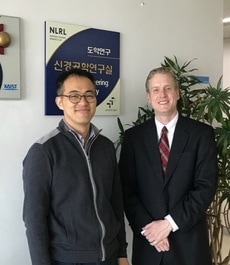
Dr. Rudi Scharnweber (MD/PhD, Department of Neurosurgery, UCLA), who studied axon guidance and local protein synthesis in axon, visited us and gave a talk on engineering of immunotherapy for the treatment of malignant glioma. Dr. Scharnweber studied with Prof. Nam in University of Urbana-Champaing in 2001 - 2006.
Feasibility Study of Extended-gate Type Silicon Nanowire Field-Effect Transistors for Neural Recording Honggi Kang, J.Y. Kim, Y.K. Choi*, Y. Nam* This is Dr. Kang's first publication since he joined NEL. We collaborated with Prof. Yang-Kyu Choi's lab to study the performance of silicon nanowire FET transistor for a novel sensing device of neural signals. The device was fabricated from Prof. Choi's lab by Dr. Kim and Dr. Kang did the measurement and analysis in terms of neural recordings. Unlike previous related works, we tried to compare the signal-to-noise ratio of the SiNW-FET with conventional metal micro electrodes. 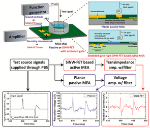 Source: Kang et al., Sensors 2017 (link)
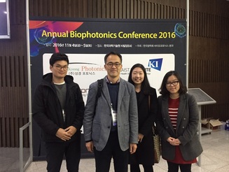
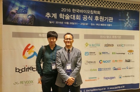
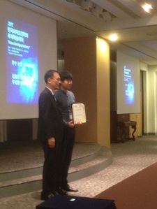
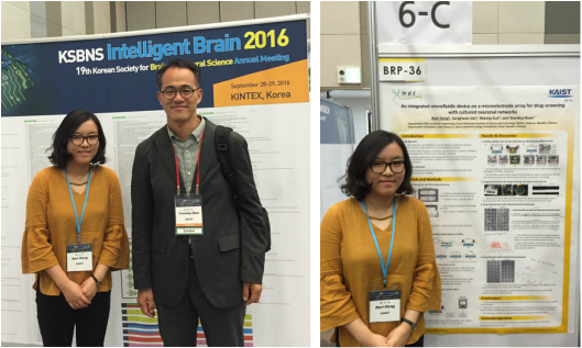
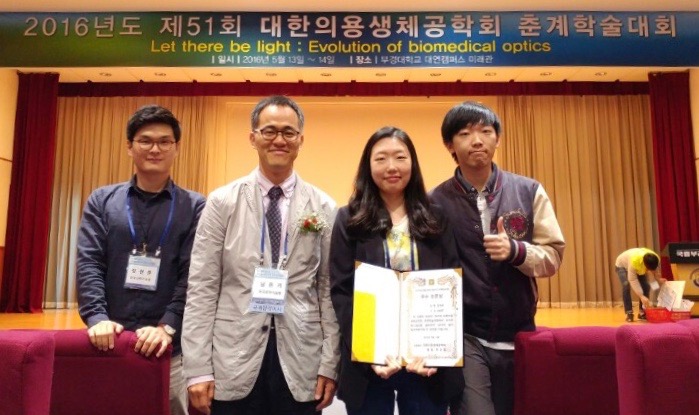
Sung-hoon Joo received Best Presentation Award.Prof. Bruce C. Wheeler (UC San Diego, Bioengineering, link), who is the pioneer of micro electrode array technology, visited us and gave a inspirational talk on current trends in biomedical engineering field. Prof. Wheeler served as the President of IEEE Engineering in Medicine and Biology Society (EMBS) in 2012 - 2013. 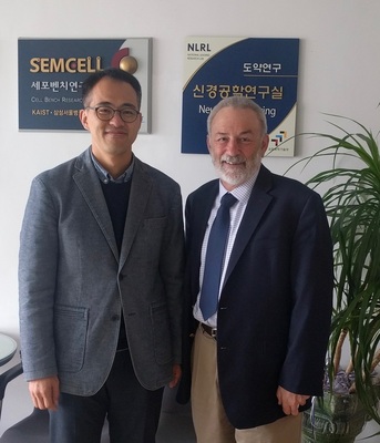 Nari Hong received Global PhD Fellowship from NRF
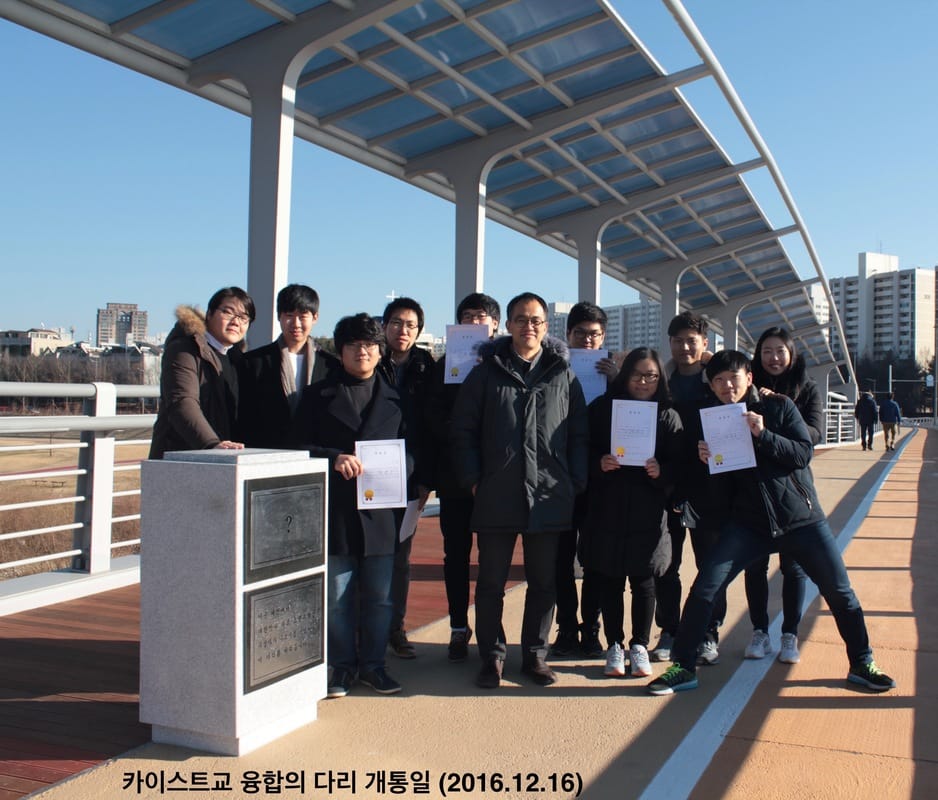
박사과정 홍나리 학생이 한국연구재단에서 선정하는 2016년 글로벌박사양성사업의 수여 대상자로 선정되었습니다. 이 사업은 국내 우수 박사과정 학생 200여명을 대상으로 3년 간 장학금을 수여합니다. 축하합니다. Characterization of axonal spikes in cultured neuronal networks using microelectrode arrays and micro channel devices
N. Hong, S. Joo, Y. Nam* This is Nari Hong's first publication reporting axonal spike recordings from dual-chamber micro channel devices which has been popular platform to study interconnected neuronal networks in vitro. Unlike other studies based on similar devices, Nari focused on characterizing axonal spikes recorded in micro-channels over a month period: signal-to-noise ratio(SNR), spike detection efficiency, axonal conduction velocity. Compared to conventional MEA recordings, we found that axonal spikes could be detected earlier in micro-channels, and conduction velocity increased over the maturation of neuronal cultures. (link) Cell-type dependent effect of surface-patterned microdot arrays on neuronal growth M. J. Jang, W. R. Kim, S. Joo, J. R. Ryu, E. Lee, Y. Nam*, W. Sun* This work is the fourth paper based on the collaboration with Prof. Woong Sun's Lab (고려대학교 의과대학 선웅 교수, Korea University College of Medicine). This is a sequel of our micro-dot array paper (Kim W, Jang MJ et al., Lab Chip 2014). In this work, Dr. Jang and Dr. Kim let the work to investigate the effect of the surface micro-dot array patterns on the growth of mouse spinal interneuron, mouse hippocampal neurons, and rat hippocampal neurons. While mouse hippocampal neurons showed no significantly different growth on control and patterned substrates, we found the microdot arrays had different effects on early neuronal growth depending on the cell type; spinal interneurons tended to grow faster in length, whereas hippocampal neurons tended to form more axon collateral branches in response to the microdot arrays. Although there was a similar trend in the neurite length and branch number of both neurons changed across the microdot arrays with the expanded range of size and spacing, the dominant responses of each neuron, neurite elongation of mouse spinal interneurons and branching augmentation of rat hippocampal neurons were still preserved. Therefore, our results demonstrate that the same design of micropatterns could cause different neuronal growth results, raising an intriguing issue of considering cell types in neural interface designs. 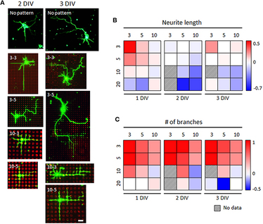 Synaptic compartmentalization by micropatterned masking of a surface adhesive cue in cultured neurons
Jae Ryun Ryu, Min Jee Jang, Youhwa Jo, Sunghoon Joo, Do Hoon Lee, Byung Yang Lee, Yoonkey Nam*, Woong Sun* This work is the third paper based on the collaboration with Prof. Woong Sun's Lab (고려대학교 의과대학 선웅 교수, Korea University College of Medicine). In this work, Dr. JR Ryu in Sun's Lab lead the project and our former member Dr. MJ Jang and PhD student S Joo closely worked to make things happen. Using our PDMS micro-stamps, we found a way to control synapse formation ('synapse focus chip') using primary hippocampal neurons. We fabricated a negative dot array pattern by coating the entire surface with poly-l-lysine (PLL) and subsequent microcontact printing of 1) substrates which mask positive charge of PLL (Fc, BSA and laminin), or 2) a chemorepulsive protein (Semaphorin 3F-Fc). By combination of physical and biological features of these repulsive substrates, functional synapses were robustly concentrated in the PLL-coated dots. (Link) KAIST, 빛과 열로 신경세포 활성 억제 성공 빛과 열로 신경세포 활성을 억제할 수 있는 길이 열렸다. KAIST는 남윤기·박지호 바이오 및 뇌공학과 교수 연구팀이 신경세포 활성을 억제할 수 있는 새로운 플랫폼을 개발했다고 30일 밝혔다. 뇌기능 연구와 뇌질환 치료에 활용될 것으로 기대된다. 연구결과는 최근 나노분야 국제학술지 `에이시에스 나노(ACS Nano)` 온라인판에 게재됐다. 연구팀은 근적외선을 선택적으로 흡수하는 금 나노막대를 합성한 후 생체 친화성을 갖는 중합체(polymer)로 코팅해 신경세포 칩 표면에 결합했다. 신경세포 칩 상 금속 전극은 금 나노막대가 결합한 후에도 전기적 특성이 변하지 않아 신경세포 활성 측정에 적합하다. 연구팀은 전기적으로 신경세포 활성을 측정하는 동시에 광열 자극으로 신경세포 활성을 억제한다는 것을 확인했다. 이 기술은 유전자 조작 없이도 빛으로 활성 조절이 가능하다. 남윤기 교수는 “나노입자와 신경세포를 결합해 새로운 자극 플랫폼을 제시했다”며 “기존 전기적 신경 시스템을 활용하는 동시에 광열 자극으로 신경세포 활성을 자유롭게 억제할 수 있다”고 말했다. KAIST 언론보도 press release (link) 전자신문 (link) (pdf) 동아사이언스 (link) 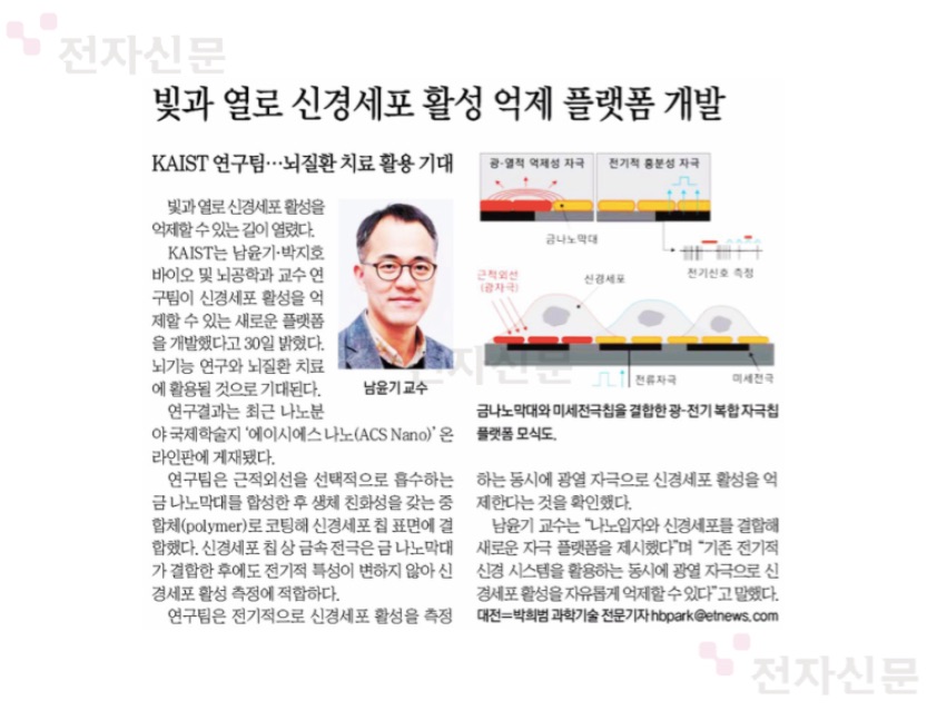 Electro-optical Neural Platform Integrated with Nanoplasmonic Inhibition Interface
Sangjin Yoo, Raeyoung Kim, Ji-Ho Park*, Yoonkey Nam* This is our second paper on optical neural inhibition technique based on gold-nanorods and near-infrared light. This work was led by Sangjin who was co-advised by Prof. Nam and Prof. Ji-Ho Park. Gold-nanorod was integrated with planar-type micro electrode array so that both electrical recording/stimulation and optical inhibition were possible in one platform. (Link) Axon-First Neuritogenesis on Vertical Nanowires
Kyungtae Kang, Yi-Seul Park, Matthew Park, Min Jee Jang, Seong-Min Kim, Juno Lee, Ji Yu Choi, Da Hee Jung, Young-Tae Chang, Myung-Han Yoon*, Jin Seok Lee*, Yoonkey Nam*, and Insung S. Choi* This is a work by our former member Kyungtae Kang who collaborated with Prof. Jin Seok Lee's lab (숙명여자대학교 화학과). Prof's Lee's lab fabricated vertically-grown silicon nanowires (vg-SiNWs) on silicon wafers and we studied the growth of cultured rat hippocampal neurons on vg-SiNW arrays. A long major neurite (axon) was formed first in neurons cultivated on vg-SiNW substrates, while neurons on conventional glass-type substrates first get short minor neurites from lamellopodia. Interestingly, cell shapes resembled those found in developing brain, which indicated the role and in-vivo like microenvironment by vg-SiNWs. (link) |
Categories
All
Archives
April 2024
|


 RSS Feed
|
Korea Advanced Institute of Science and Technology (KAIST)
291 Daehak-ro, Yuseong-gu Daejeon 34141, Republic of Korea https://www.kaist.ac.kr Phone: +82-42-350-5362 |
|