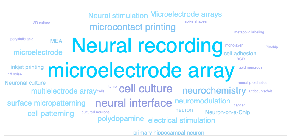
Current research area includes
- Microelectrode Array technology
- Thermoplasmonic neural interfaces
- Cell patterning techniques
- Bio-fucntionalization of neural interfaces
- Neuromodulation techniques
- Multichannel neural information analysis
- Neurons-on-Nanomaterials
MEA technology
We design a novel planar microelectrode array (MEA) system that can be used to grow live neurons and record neural signals from multiple neurons simultaneously. Electrical and optical stimulation and recording techniques are combined with the MEA platform to investigate neural circuits in vitro. 60 to 256 channel MEAs have been successfully designed with nanostructure microelectrode electrodes (e.g. conductive polymer PEDOT:PSS).
We design a novel planar microelectrode array (MEA) system that can be used to grow live neurons and record neural signals from multiple neurons simultaneously. Electrical and optical stimulation and recording techniques are combined with the MEA platform to investigate neural circuits in vitro. 60 to 256 channel MEAs have been successfully designed with nanostructure microelectrode electrodes (e.g. conductive polymer PEDOT:PSS).
- Compact 256-channel multi-well microelectrode array system for in vitro neuropharmacology test, Lab Chip. 2020. (link)
- Polydopamine-doped conductive polymer microelectrodes for neural recording and stimulation, J Neurosci Methods. 2019. (link)
- Electrochemical layer-by-layer approach to fabricate mechanically stable platinum black microelectrodes using a mussel-inspired polydopamine adhesive, J Neural Eng. 2015. (Link)
- Gold nanograin microelectrodes for neuroelectronic interfaces, Biotechnol J, 2013. (Link)
- Surface-modified microelectrode array with flake nanostructure for neural recording and stimulation, Nanotechnology, 2010. (Link)
- In Vitro Neural Recording by Microelectrode Arrays, book chapter in Stretchable Bioelectronics for Medical Devices and Systems, Springer. (link)
- Recent trends in microelectrode array technology for in vitro neural interface platform, Biomed Eng Lett. 2014. (Link)
- Material considerations for in vitro neural interface technology, MRS Bulletin 2012. (Link)
- In vitro microelectrode array technology and neural recordings, Crit Rev Biomed Eng. 2011. (Link)
Nano-Neurotechnology: Thermoplasmonic neural interfaces
We use nano-transducers (e.g. gold nanorods) to design photothermal neural interfaces for controlling neural spiking activity. Photothermal conversion was successfully implemented to induce reversible inhibition of spiking activity for more than tens of minutes. Using the novel inhibition effect, the regulation of spiking activity will be possible for hyperactive neural circuits in the future.
We use nano-transducers (e.g. gold nanorods) to design photothermal neural interfaces for controlling neural spiking activity. Photothermal conversion was successfully implemented to induce reversible inhibition of spiking activity for more than tens of minutes. Using the novel inhibition effect, the regulation of spiking activity will be possible for hyperactive neural circuits in the future.
- Thermoplasmonic neural chip platform for in situ manipulation of neuronal connections in vitro, Nature Communications. 2020 (link)
- Single-Cell Photothermal Neuromodulation for Functional Mapping of Neural Networks. ACS Nano. 2019 (link)
- Inkjet-Printed Bio-Functional Thermo-Plasmonic Interfaces for Patterned Neuromodulation. ACS Nano, 2018. (link)
- Gold nanostar-mediated neural activity control using plasmonic photothermal effects. Biomaterials. 2017. (link) [KAIST Breakthroughs: (link)]
- Digital Micromirror based Near-infrared Illumination System for Plasmonic Photothermal Neuromodulation. Biomedical Optics Express 2017. (Link)
- Electro-optical Neural Platform Integrated with Nanoplasmonic Inhibition Interface, ACS Nano. 2016. (Link)
- Photothermal Inhibition of Neural Activity with Near-Infrared-Sensitive Nanotransducers, ACS Nano. 2014.(Link)
2D-3D Cell patterning techniques for the Brain-on-a-Chip technology
We want to build live biological neuronal networks in vitro with ordered structures. We believe that the cultured neuronal network is a good model system to study basic principles of learning and memory at a network level. Micro-contact printing technique is the major method to print biomolecules that can either promote or prohibit neuronal growth. Surface chemical cues and topographical cues are the design rules for the guided neural growth in vitro. We are interested in discovering new physical and chemical guidance cues that lead to neuronal polarization and migration.
We want to build live biological neuronal networks in vitro with ordered structures. We believe that the cultured neuronal network is a good model system to study basic principles of learning and memory at a network level. Micro-contact printing technique is the major method to print biomolecules that can either promote or prohibit neuronal growth. Surface chemical cues and topographical cues are the design rules for the guided neural growth in vitro. We are interested in discovering new physical and chemical guidance cues that lead to neuronal polarization and migration.
- Design and fabrication of miniaturized neuronal circuits on microelectrode arrays using agarose hydrogel micro-molding technique, BioChip Journal, 2018. (link)
- Stimuli-responsive neuronal networking via removable alginate masks, Advanced Biosystems, 2018. (link)
- Effects of ECM protein micropatterns on the migration and differentiation of adult neural stem cells, Scientific Reports, 2015. (Link)
- In vitro neurite guidance effects induced by polylysine pin-stripe micropatterns with polylysine background, J Biomed Mater Res A, 2015. (Link)
- Surface-printed microdot array chips for the quantification of axonal collateral branching of a single neuron in vitro, Lab Chip. 2014. (Link)
- Geometric effect of cell adhesive polygonal micropatterns on neuritogenesis and axon guidance, J Neural Eng, 2012. (Link)
- Agarose microwell based neuronal micro-circuit arrays on microelectrode arrays for high throughput drug testing, Lab Chip, 2009. (Link)
Bio-functionalization of neural interfaces
In order to grow neurons in vitro, we design the cell-surface interface using chemical functionalization approaches and surface micro patterning. In addition, we are actively searching for a simple and effective cell-adhesive or -repellent substrates. So far, we have pioneered in using a mussel-inspired polymer coating ('polydopamine') in MEA for covalently linking cell-adhesive biomolecules on metal and insulator.
In order to grow neurons in vitro, we design the cell-surface interface using chemical functionalization approaches and surface micro patterning. In addition, we are actively searching for a simple and effective cell-adhesive or -repellent substrates. So far, we have pioneered in using a mussel-inspired polymer coating ('polydopamine') in MEA for covalently linking cell-adhesive biomolecules on metal and insulator.
- Electrochemically driven, electrode-addressable formation of functionalized polydopamine films for neural interfaces, Angew Chem Int Ed Engl, 2012. (Link)
- A biofunctionalization scheme for neural interfaces using polydopamine polymer, Biomaterials, 2011. (Link)
- Agarose-assisted micro-contact printing for high-quality biomolecular micro-patterns, Macromolecular Bioscience, 2015. (Link)
- Aqueous micro-contact printing of cell-adhesive biomolecules for patterning neuronal cell cultures, BioChip J, 2012. (Link)
- Neurons on Parafilm: versatile elastic substrates for neuronal cell cultures, J Neurosci Methods, 2012. (Link)
Multichannel neural information analysis
We are interested in the relation between network structures and information processing in neuronal network. Microelectrode array and optical imaging are two major platforms to access multiple neurons simultaneously. Recently, we have developed a large-scale optophysiological data processing software, NeuroCa (link), that can efficiently handle a big data problem in neuroinformatics.
We are interested in the relation between network structures and information processing in neuronal network. Microelectrode array and optical imaging are two major platforms to access multiple neurons simultaneously. Recently, we have developed a large-scale optophysiological data processing software, NeuroCa (link), that can efficiently handle a big data problem in neuroinformatics.
- Connectivity and network burst properties of in-vitro neuronal networks induced by a clustered structure with alginate hydrogel patterning, Biomedical Eng. Lett. (link) (Matlab code)
- NeuroCa: Integrated framework for systematic analysis of spatio-temporal neuronal activity patterns from large-scale optical recording data, Neurophoton., 2(3), 035003 (2015). (Link) (NeuroCa)
- Inference of combinatorial neuronal synchrony with Bayesian networks, J Neurosci Methods, 2010. (Link)
- Systematic analysis of synchronized oscillatory neuronal networks reveals an enrichment for coupled direct and indirect feedback motifs, Bioinformatics, 2009. (Link)
Neurons-on-Nanomaterials
We study nano-scale neural interfaces by growing primary neurons on various nano-topographical surfaces including carbon nano-tubes, anodised aluminium oxides (AAO), self-assembled silica nanobeads, and vertically grown silicon nanowire. We focused on effects of surface properties on the early neuronal developments such as growth cone formation, axon formation, neuritogensis, etc. So far, we found that nano-scale topographical features affect neurite formation and neuronal maturation.
We study nano-scale neural interfaces by growing primary neurons on various nano-topographical surfaces including carbon nano-tubes, anodised aluminium oxides (AAO), self-assembled silica nanobeads, and vertically grown silicon nanowire. We focused on effects of surface properties on the early neuronal developments such as growth cone formation, axon formation, neuritogensis, etc. So far, we found that nano-scale topographical features affect neurite formation and neuronal maturation.
- Axon-First Neuritogenesis on Vertical Nanowires, Nano Lett., (Link)
- Cytoskeletal Actin Dynamics are Involved in Pitch-Dependent Neurite Outgrowth on Bead Monolayers, Angew Chem Int Ed Engl. 2014. (Link)
- In vitro developmental acceleration of hippocampal neurons on nanostructures of self-assembled silica beads in filopodium-size ranges, Angew Chem Int Ed Engl, 2012. (Link)
- Pitch-dependent acceleration of neurite outgrowth on nanostructured anodized aluminum oxide substrates, Angew Chem Int Ed Engl, 2010. (Link)
- Directional neurite growth using carbon nanotube patterned substrates as a biomimetic cue, Nanotechnology, 2010. (Link)
* Funding sources: KAIST, National Research Foundation, Samsung Science & Technology Foundation