|
Characterization of axonal spikes in cultured neuronal networks using microelectrode arrays and micro channel devices
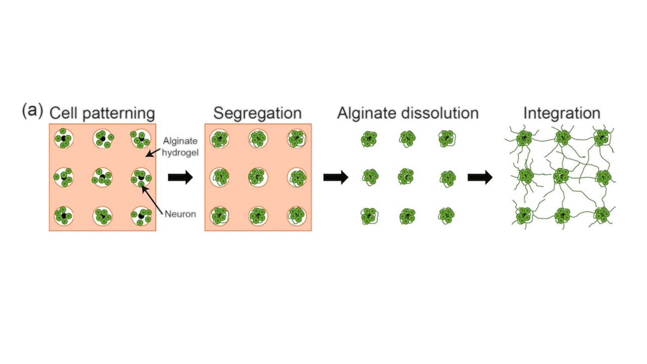
N. Hong, S. Joo, Y. Nam* This is Nari Hong's first publication reporting axonal spike recordings from dual-chamber micro channel devices which has been popular platform to study interconnected neuronal networks in vitro. Unlike other studies based on similar devices, Nari focused on characterizing axonal spikes recorded in micro-channels over a month period: signal-to-noise ratio(SNR), spike detection efficiency, axonal conduction velocity. Compared to conventional MEA recordings, we found that axonal spikes could be detected earlier in micro-channels, and conduction velocity increased over the maturation of neuronal cultures. (link) Cell-type dependent effect of surface-patterned microdot arrays on neuronal growth
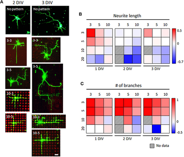
M. J. Jang, W. R. Kim, S. Joo, J. R. Ryu, E. Lee, Y. Nam*, W. Sun* This work is the fourth paper based on the collaboration with Prof. Woong Sun's Lab (고려대학교 의과대학 선웅 교수, Korea University College of Medicine). This is a sequel of our micro-dot array paper (Kim W, Jang MJ et al., Lab Chip 2014). In this work, Dr. Jang and Dr. Kim let the work to investigate the effect of the surface micro-dot array patterns on the growth of mouse spinal interneuron, mouse hippocampal neurons, and rat hippocampal neurons. While mouse hippocampal neurons showed no significantly different growth on control and patterned substrates, we found the microdot arrays had different effects on early neuronal growth depending on the cell type; spinal interneurons tended to grow faster in length, whereas hippocampal neurons tended to form more axon collateral branches in response to the microdot arrays. Although there was a similar trend in the neurite length and branch number of both neurons changed across the microdot arrays with the expanded range of size and spacing, the dominant responses of each neuron, neurite elongation of mouse spinal interneurons and branching augmentation of rat hippocampal neurons were still preserved. Therefore, our results demonstrate that the same design of micropatterns could cause different neuronal growth results, raising an intriguing issue of considering cell types in neural interface designs. |
Categories
All
Archives
December 2023
|
|
Korea Advanced Institute of Science and Technology (KAIST)
291 Daehak-ro, Yuseong-gu Daejeon 34141, Republic of Korea https://www.kaist.ac.kr Phone: +82-42-350-5362 |


 RSS Feed
