|
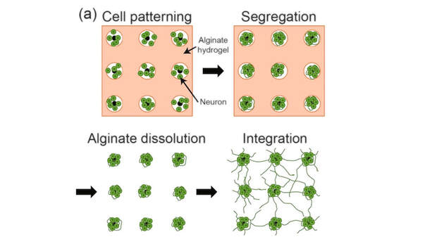
이형섭 박사 (2023. 8월 졸업, 현, 삼성전자)의 박사학위 논문 일부가 국제 학술지 Biomedical Engineering Letters (2022 I/F:4.6) 에 게재되었습니다. 이 연구는 우리 실험실에서 개발한 군집형 신경네트웍 배양 모델을 시험관 조건에서 약 1달 간 배양하면서 신경세포 군집들 간의 생물학적 네트웍 형성 과정을 다채널 미세전극칩으로 측정하고, 네트웍의 구조와 기능과의 관계를 시계열 특성, 시공간 패턴, 모티브 등을 분석한 논문입니다. Hyungsub's(2023. 08 graduate, now Samsung Electronics) PhD work has been published in Biomedical Engineering Letters. In this study, he investigated the development of in vitro clustered neuronal networks using the microelectrode array recording technology. Multichannel spike train analysis was used to investigate the functional connectivity and motifs emerging from the network. (Source)
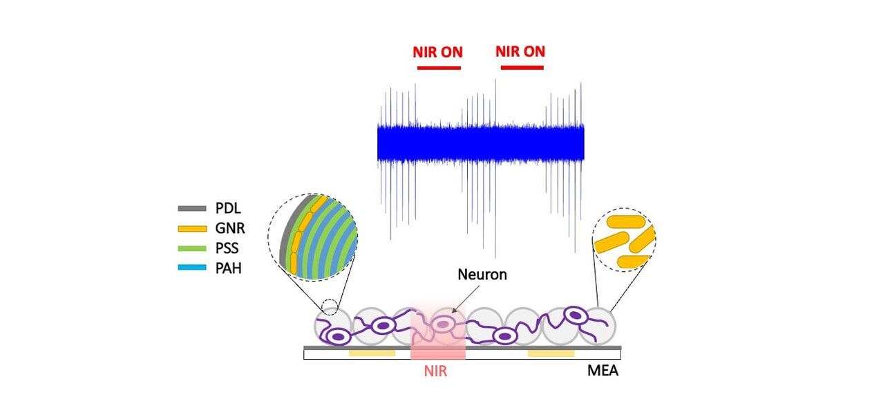
H Lee, GS Lee, Y Nam*, "Connectivity and network burst properties of in-vitro neuronal networks induced by a clustered structure with alginate hydrogel patterning", Biomedical Engineering Letters (link) (Matlab code) 우리 연구실 석사과정 교환학생 Andrea Andolfi (Univ of Genoa, Italy)이 수행한 3차원 광열 신경자극 플랫폼에 관한 연구가 국제저명학술지 BioChip Journal (I/F 4.229) 에 게재되었습니다. 이 연구는 미세 유리구슬 (microglass bead) 에 Layer-by-layer 코팅 기법을 이용하여 금나노막대 기반 3차원 광열신경자극 인터페이스를 제작하는 기법에 대한 보고입니다. 석사졸업생 장현수 님이 이 연구에 참가하였습니다.
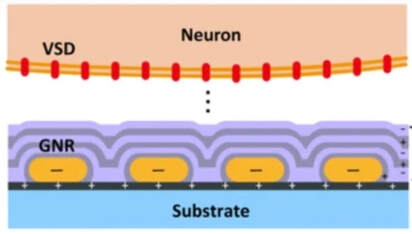
Figure source: Andolfi, A., Jang, H., Martinoia, S. et al. Thermoplasmonic Scaffold Design for the Modulation of Neural Activity in Three-Dimensional Neuronal Cultures. BioChip J (2022). https://doi.org/10.1007/s13206-022-00082-z Fabrication of a Nanoplasmonic Chip to Enhance Neuron Membrane Potential Imaging by Metal-Enhanced Fluorescence Effect
Dr. Raeyoung Kim's PhD work on the study of nanoplasmonics based metal-enhanced fluorescence effect on voltage-sensitive dye imaging was published in BioChip Journal. Congratulations! Inkjet-Printed Bio-Functional Thermo-Plasmonic Interfaces for Patterned Neuromodulation
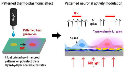
Hongki Kang, Gu-Haeng Lee, Hyunjun Jung, Jee Woong Lee, Yoonkey Nam* Based on our previous contributions on nanoplamonic neural interface platform (2014, 2016), we made another big step toward the design of plasmonic neural interface devices by adopting state-of-the-art inkjet printing technology. We propose an innovative, but also practically very efficient, approach to fabricate sophisticated thermo-plasmonic micropatterns on any type of substrates (rigid/flexible, hydrophobic/hydrophilic) using NIR-sensitive gold nanorods. We found that integrating polyelectrolyte layer-by-layer assembly with inkjet printing can provide excellent pattern fidelity and uniformity of micro-patterned thermo-plasmonic nanoparticles on various substrates for neuromodulation. With this printing process, we demonstrated that thermo-plasmonic heat sources can be fabricated over very large area (a few cm scale) with great design flexibility. Moreover, the bio-functionality of this high quality thermo-plasmonic interface was also confirmed by selectively modulating electrical activity of in vitro neuronal networks. Inkjet-printed multi-wavelength thermo-plasmonic images for anti-counterfeiting applications
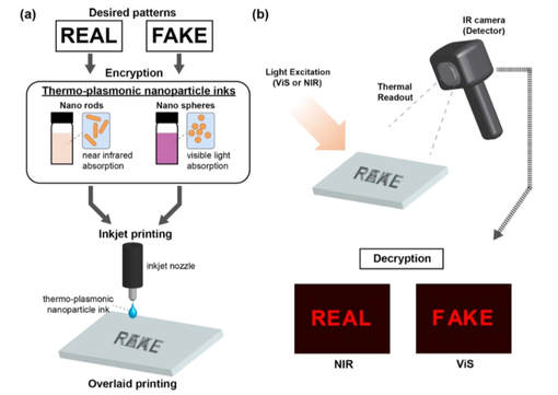
Hongki Kang, Jee Woong Lee, Yoonkey Nam* This work was led by Dr. Kang in collaboration with Jee Woong. We used two different types of gold nano-particles tuned at different wavelengths (ViS vs. NIR) so that nano-particles can generate heat at specific light. Using an optimized inkjet printing for gold nanoparticles, we we were able to print desired patterns or codes on substrates and the patterns can be decoded through wavelength specific thermal images. We demonstrated that this platform can be applied for anti-counterfeiting applications. Gold nanostar-mediated neural activity control using plasmonic photothermal effects
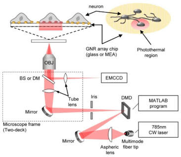
Jee Woong Lee, Hyunjun Jung, Hui Hun Cho, Jung Heon Lee, Yoonkey Nam* This work was led by Jee Woong in collaboration with Prof. Jung Heon Lee's lab (link) at Sungkyunkwan University. We used gold nanostar shaped nanoparticles to induce photothermal effects for neuromodulation. Unlike gold nanorods that are mainly used for phototheraml effects, our gold nano stars were synthesized by seedless methods under completely biocompatibile environment. We showed that gold nanostars were effective in inducing photothermal neural inhibition from single cell level to network level. HJ jung also contributed to this work with his DMD-based phototheraml patterning system for the single cell suppression experiment. (link) Digital Micromirror based Near-infrared Illumination System for Plasmonic Photothermal Neuromodulation
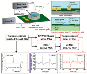
Hyunjun Jung, H. Kang, Y. Nam This is Hyunjun Jung's first paper on his digital micro mirror system for NIR neural suppression experiments. In this paper, Jung reports the patterned photothermal stimulation of neuronal networks with plasmonic gold nanorod MEA chips. Feasibility Study of Extended-gate Type Silicon Nanowire Field-Effect Transistors for Neural Recording Honggi Kang, J.Y. Kim, Y.K. Choi*, Y. Nam* This is Dr. Kang's first publication since he joined NEL. We collaborated with Prof. Yang-Kyu Choi's lab to study the performance of silicon nanowire FET transistor for a novel sensing device of neural signals. The device was fabricated from Prof. Choi's lab by Dr. Kim and Dr. Kang did the measurement and analysis in terms of neural recordings. Unlike previous related works, we tried to compare the signal-to-noise ratio of the SiNW-FET with conventional metal micro electrodes. Source: Kang et al., Sensors 2017 (link)
Characterization of axonal spikes in cultured neuronal networks using microelectrode arrays and micro channel devices
N. Hong, S. Joo, Y. Nam* This is Nari Hong's first publication reporting axonal spike recordings from dual-chamber micro channel devices which has been popular platform to study interconnected neuronal networks in vitro. Unlike other studies based on similar devices, Nari focused on characterizing axonal spikes recorded in micro-channels over a month period: signal-to-noise ratio(SNR), spike detection efficiency, axonal conduction velocity. Compared to conventional MEA recordings, we found that axonal spikes could be detected earlier in micro-channels, and conduction velocity increased over the maturation of neuronal cultures. (link) Cell-type dependent effect of surface-patterned microdot arrays on neuronal growth
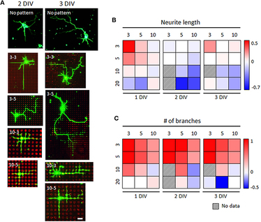
M. J. Jang, W. R. Kim, S. Joo, J. R. Ryu, E. Lee, Y. Nam*, W. Sun* This work is the fourth paper based on the collaboration with Prof. Woong Sun's Lab (고려대학교 의과대학 선웅 교수, Korea University College of Medicine). This is a sequel of our micro-dot array paper (Kim W, Jang MJ et al., Lab Chip 2014). In this work, Dr. Jang and Dr. Kim let the work to investigate the effect of the surface micro-dot array patterns on the growth of mouse spinal interneuron, mouse hippocampal neurons, and rat hippocampal neurons. While mouse hippocampal neurons showed no significantly different growth on control and patterned substrates, we found the microdot arrays had different effects on early neuronal growth depending on the cell type; spinal interneurons tended to grow faster in length, whereas hippocampal neurons tended to form more axon collateral branches in response to the microdot arrays. Although there was a similar trend in the neurite length and branch number of both neurons changed across the microdot arrays with the expanded range of size and spacing, the dominant responses of each neuron, neurite elongation of mouse spinal interneurons and branching augmentation of rat hippocampal neurons were still preserved. Therefore, our results demonstrate that the same design of micropatterns could cause different neuronal growth results, raising an intriguing issue of considering cell types in neural interface designs. Synaptic compartmentalization by micropatterned masking of a surface adhesive cue in cultured neurons
Jae Ryun Ryu, Min Jee Jang, Youhwa Jo, Sunghoon Joo, Do Hoon Lee, Byung Yang Lee, Yoonkey Nam*, Woong Sun* This work is the third paper based on the collaboration with Prof. Woong Sun's Lab (고려대학교 의과대학 선웅 교수, Korea University College of Medicine). In this work, Dr. JR Ryu in Sun's Lab lead the project and our former member Dr. MJ Jang and PhD student S Joo closely worked to make things happen. Using our PDMS micro-stamps, we found a way to control synapse formation ('synapse focus chip') using primary hippocampal neurons. We fabricated a negative dot array pattern by coating the entire surface with poly-l-lysine (PLL) and subsequent microcontact printing of 1) substrates which mask positive charge of PLL (Fc, BSA and laminin), or 2) a chemorepulsive protein (Semaphorin 3F-Fc). By combination of physical and biological features of these repulsive substrates, functional synapses were robustly concentrated in the PLL-coated dots. (Link) Electro-optical Neural Platform Integrated with Nanoplasmonic Inhibition Interface
Sangjin Yoo, Raeyoung Kim, Ji-Ho Park*, Yoonkey Nam* This is our second paper on optical neural inhibition technique based on gold-nanorods and near-infrared light. This work was led by Sangjin who was co-advised by Prof. Nam and Prof. Ji-Ho Park. Gold-nanorod was integrated with planar-type micro electrode array so that both electrical recording/stimulation and optical inhibition were possible in one platform. (Link) Axon-First Neuritogenesis on Vertical Nanowires
Kyungtae Kang, Yi-Seul Park, Matthew Park, Min Jee Jang, Seong-Min Kim, Juno Lee, Ji Yu Choi, Da Hee Jung, Young-Tae Chang, Myung-Han Yoon*, Jin Seok Lee*, Yoonkey Nam*, and Insung S. Choi* This is a work by our former member Kyungtae Kang who collaborated with Prof. Jin Seok Lee's lab (숙명여자대학교 화학과). Prof's Lee's lab fabricated vertically-grown silicon nanowires (vg-SiNWs) on silicon wafers and we studied the growth of cultured rat hippocampal neurons on vg-SiNW arrays. A long major neurite (axon) was formed first in neurons cultivated on vg-SiNW substrates, while neurons on conventional glass-type substrates first get short minor neurites from lamellopodia. Interestingly, cell shapes resembled those found in developing brain, which indicated the role and in-vivo like microenvironment by vg-SiNWs. (link) Effects of ECM protein micropatterns on the migration and differentiation of adult neural stem cells
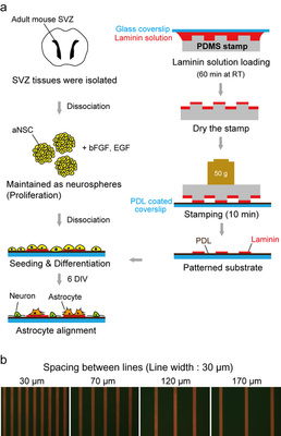
Sunghoon Joo, Joo Yeon Kim, Eunsoo Lee, Nari Hong, Woong Sun & Yoonkey Nam This is an outcome of our on-going collaboration with Prof. Woong Sun's lab in Korea University (고려대학교 해부학 교실, 선웅 교수). In this work, Sunghoon (KAIST, NEL) and Joo Yeon (Korea University College of Medicine) applied soft-lithographic technique to design simple and reproducible laminin (LN)-polylysine cell culture substrates and investigated how aNSCs respond to the various spatial distribution of laminin, one of ECM proteins enriched in the aNSC niche. They found that aNSC preferred to migrate and attach to LN stripes, and aNSC-derived neurons and astrocytes showed significant difference in motility towards LN stripes. By changing the spacing of LN stripes, they discovered a new way of separating neurons and astrocytes differentiated from adult neural stem cells. To the best of our knowledge, this is the first time to investigate the differential cellular responses of aNSCs on ECM protein (LN) and cell adhesive synthetic polymer (PDL) using surface micropatterns. NeuroCa: Integrated framework for systematic analysis of spatio-temporal neuronal activity patterns from large-scale optical recording data
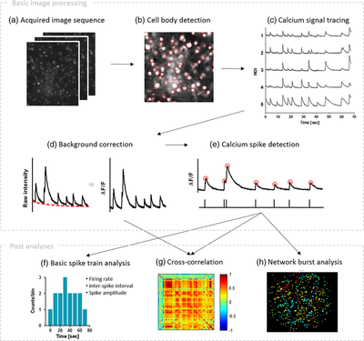
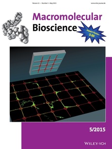
Minjee Jang and Yoonkey Nam In this work, Minjee developed a MATLAB-based toolbox, named NeuroCa, for the automated processing and quantitative analysis of large-scale calcium imaging data. This tool includes several computational algorithms to extract the calcium spike trains of individual neurons from the calcium imaging data in an automatic fashion. Two algorithms were developed to decompose the imaging data into the activity of individual cells and subsequently detect calcium spikes from each neuronal signal. Applying our method to dense networks in dissociated cultures, we were able to obtain the calcium spike trains of ∼1000 neurons in a few minutes. Further analyses using these data permitted the quantification of neuronal responses to chemical stimuli as well as functional mapping of spatiotemporal patterns in neuronal firing within the spontaneous, synchronous activity of a large network. These results demonstrate that our method not only automates time-consuming, labor-intensive tasks in the analysis of neural data obtained using optical recording techniques but also provides a systematic way to visualize and quantify the collective dynamics of a network in terms of its cellular elements. For more information please go to the NeuroCa webpage (link) Minjee Jang's work on agarose-assisted microstamping method was selected as the cover article. Dae-Jeong Kim helped to design the cover image. Agarose-assisted micro-contact printing uses a stamp covered with agarose thin film, enabling the effective patterning of biomolecules to guide neuronal growth on the substrate. Various patterns including dots with a few micrometers in diameter are also available to be patterned using the method presented by M. J. Jang and Y. Nam on page 613. (Cover designed by M. J. Jang and D. J. Kim.) Source: Jang et al., Macro. Bioscience 2015 (link)
Electrochemical layer-by-layer approach to fabricate mechanically stable platinum black microelectrodes using a mussel-inspired polydopamine adhesive
Raeyoung Kim and Yoonkey Nam In this work, we applied a mussel-inspired polydopamine chemistry to fabricate mechanically stable neural microelectrodes (platinum black electrodes). The main motivation was to show that polydopamine can serve as a molecular adhesive layer to stablize platinum black nanostructures on gold film. Using our previously developed electrochemical deposition of polydopamine layers, we successfully developed a platinum black hybrid micro-structures that had superior mechanical properties. (Link) Photothermal Inhibition of Neural Activity with Near-Infrared-Sensitive Nanotransducers Sangjin Yoo, Soonwoo Hong, Yeonho Choi, Ji-Ho Park*, and Yoonkey Nam* This work reports a novel finding of photothermal inhibition of neuronal spiking activity using NIR sensitive gold nanorods. We showed that action potentials were effectively inhibited at a single cell level, and thus the neural spike rates can be modulated through the light-mediated thermal stimulation. Furthermore, thermo-sensitive potassium channel was involved in the inhibition effect. Sangjin Yoo, who lead this project for 3 years, has been co-advised by Prof. Ji-ho Park in our department. Prof. Y. Choi and his student Mr. Hong at Korea University also contributed in calculating the temperature profile of the gold-nanorods. Source: Yoo et al., ACS Nano 2014 (Link)
Electrochemically Driven, Electrode-Addressable Formation of Functionalized Polydopamine Films for Neural Interfaces. Kyungtae Kang, Seokyoung Lee, Raeyoung Kim, Insung S. Choi*,Yoonkey Nam,* This work reports a new method to selectively bio-functionalize neural interfaces using bioinspired adhesive film ('polydopamine'). We showed that neuron-adhesive biomolecules and polymers (polylysine, RGD peptides) can be linked on microelectrode surfaces via covalent bonds using electrochemically synthesized polydopamine film. (link)
Gold nanograin microelectrodes for neuroelectronic interfaces.
Raeyoung Kim, Nari Hong and Yoonkey Nam* This work reported a design of microelectrodes using gold nanostructures ('nanograin') for neural recording and stimulation. We developed a electrochemical deposition technique to generate a nanograin structures on metal microelectrodes and characterized metal-electrolyte interfaces using AC impedance spectroscopy and current injection tests. (link) Geometric effect of cell adhesive polygonal micropatterns on neuritogenesis and axon guidance. Min Jee Jang and Yoonkey Nam*
This work investigated the shape effect of micro-printed chemical cues on neuronal development in vitro. We found that triangle shapes can be used to control neurite outgrowth and axonal development in vitro. (Link) Aqueous micro-contact printing of cell-adhesive biomolecules for patterning neuronal cell cultures, Min Jee Jang and Yoonkey Nam*. This work shows that conventional micro-contact printing can be done under the aqueous conditions withtout compromising the minimum printable feature sizes. This method will overcome some limitations of micro-contact printings used for creating live neural circuits in vitro. (Link)
Identification of feedback loops in neural networks based on multi-step Granger causality.
Dong CY, Shin D, Joo S, Nam Y, Cho KH*. This work is a collaboration with Prof. Kwang Hyun Cho's lab to analyze synchronized neural activity from cultured neuron. Sung-hoon Joo (MS student) participated in this project by performing experiments with cultured neurons. This paper is our second paper with Prof. Cho's lab. (Link) Material considerations for in vitro neural interface technology, Yoonkey Nam Prof. Nam's new review article has been published in MRS Bulletin (June). MRS (Materais Research Society) is an internationally recognized academic society consisting of about 16,000 members world-wide. In this month, MRS Bulletin featured material issues for neural interfaces and Prof. Nam was invited to contribute an article on in vitro neural interface technology. (Link)
In Vitro Developmental Acceleration of Hippocampal Neurons on Nanostructures of Self-Assembled Silica Beads in Filopodium-Size Ranges
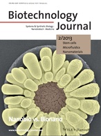
Kyungtae Kang, S Choi, HS Jang, WK Cho, Yoonkey Nam*, IS Choi*, JS Lee*. This work investigated the effect of nanotopographical scales on neurite outgrowth using silica nanobeads. The beads were synthesized by Prof. JS Lee's lab in Suk-Myung University and cell culture and analysis were performed in Neural Engineering Lab. (link to article) This work was also selected as a cover picture (link). Congratulations! |
Categories
All
Archives
December 2023
|
|
Korea Advanced Institute of Science and Technology (KAIST)
291 Daehak-ro, Yuseong-gu Daejeon 34141, Republic of Korea https://www.kaist.ac.kr Phone: +82-42-350-5362 |

 RSS Feed
